


Arm DVT
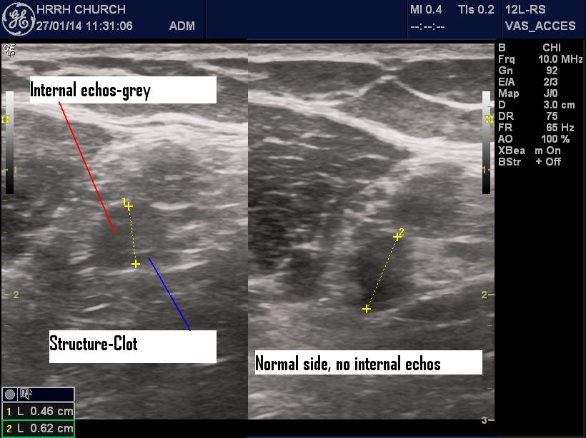
Dr. Lloyd Gordon has a case of proximal arm DVT to share. Like doing a scan for proximal leg DVT, looking for lack of vein compressibility is key but in the majority of cases if you look carefully you will see some clot in the lumen with varying amounts of echogenicity.
So while most of us learned to do DVT scans on the leg, you can certainly use the same skill to check for clots in other vessels. Just remember that in the complex patient or those with high pre-test probability of clot, trust your clinical instincts and get further investigations done if you don’t see the clot yourself.
[ed.]
This patient had a long history of arm DVT so it didn’t take a genius to figure out the diagnosis.
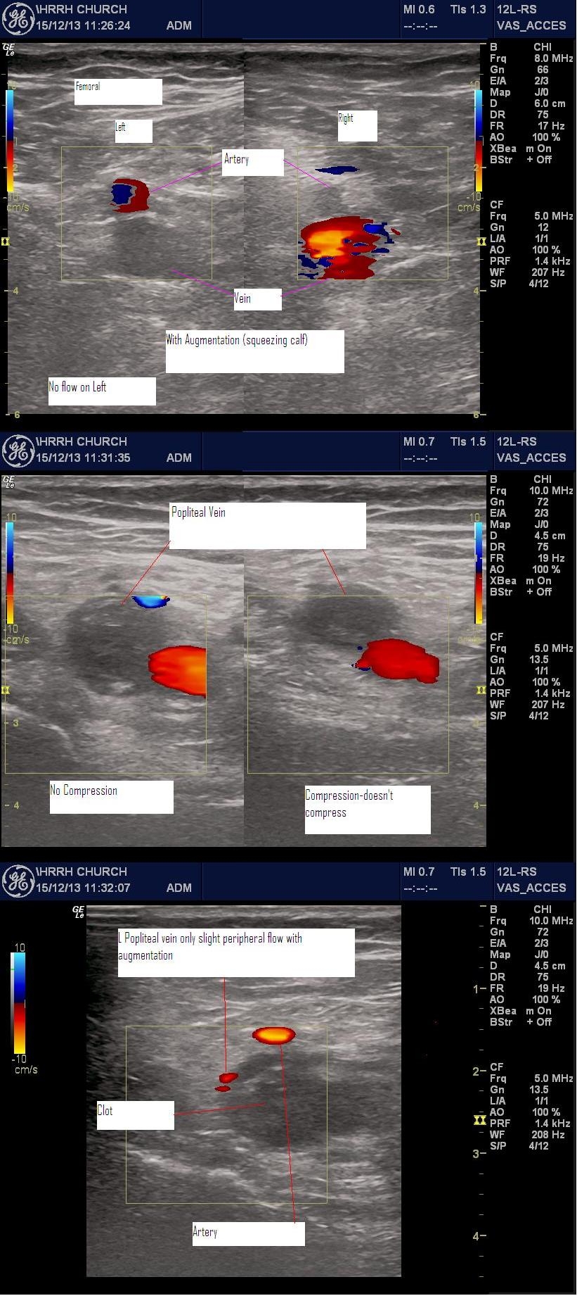
POCUS revealed normal compressibility of the brachial vein. However the affected segment changed from a normal U/S look (black, no structure) to a “Clotty” look (grey with echos suggesting texture). Flow and augmented flow were reduced in the affected segment.
